Add ~200 ml protein gel stain. (b) You may want to add a Kimwipe to the destaining solution to adsorb the dye.

Anime & Manga 4.

Selected isolates were inoculated into a sterile 50-ml conical flask containing nutrient broth and incubated in an orbital shaker (250 rpm) for 90 to 110 h at room temperature. Most widely used method of this family is the Bradford method 5 in which Coomassie blue G. Coomassie Plus Bradford Assay Kit Thermo Fisher Scientific.
 abcam coomassie stain
 Coomassie Stain Protocol The gel must be fixed prior to staining by a non-modifying, precipitation procedure such as the ethanol (or methanol)-acetic acid method used here. 3. Colloidal Coomassie staining according to Neuhoff (Electrophoresis 1988, 9, 255-262) Detection limit: 0.7 ng/mm2gel (for normal Coomassie: 20 -100 ng/mm2 gel) 1. Stock solutions: Solution A: 10% (w/v) ammonium sulphate, 2% (w/v) phosphoric acid in MilliQ water Solution B: 5% (w/v) Coomassie Brilliant Blue G-250 in MilliQ water
staining coomassie gels proteins organic without Car 7.
Coomassie blue staining 
5.1 Standard Protocol 1. 4305 Orders Completed. Caution: Use caution while performing the following steps using a microwave oven.
Coomassie blue staining - PubMed Coomassie staining is one of the simplest non-radioactive methods for visualizing proteins in gels. Staining for 16 hr allows detection of amounts <10 ng of BSA Bioz Stars score: 99/100, based on 1 PubMed citations. The detailed protocol is described below: The staining solution is prepared by mixing 100 ml of the stock solution A with 2.5 ml stock solution B.
western virus overlay blot blotting far assay coomassie protocol bio sds figure purified analysis 
10% acetic acid.


 coomassie staining g membrane igg
 120 mL . Stain: Dissolve 0.4g of Coomassie blue R350 in 200 mL of 40% (v/v) methanol in water with stirring as needed.
coomassie gel troubleshooting stain electrophoresis solution doesn problem below Destain: Enter the email address you signed up with and we'll email you a reset link. Modified GelCode Blue Coomassie Stain Reagents 1. Bio safe coomassie stain.
Stain Protocols 
$16: She helped me in last minute in a very reasonable price. Awesome 6.
coomassie staining protein 250 gels stain r250 stains differ dyes methyl function groups Colloidal Coomassie Staining Protocol Reagents: Fixing Solution: 40% methanol, 7% acetic acid 53 mL MilliQ water 40 mL methanol 7 mL acetic acid Staining Solution: 1X Brilliant Blue G-Colloidal Coomassie 800 ml MilliQ water to concentrate. *Mix by inversion.
coomassie anol bio safe coomassie stain. Submerge the gel in enough Coomassie Blue staining solution so that the gel floats freely in the tray. Shake slowly on a laboratory shaker for 30 min - 2 h. The amount of time required to stain the gel depends on the thickness of the gel. A 0.75 mm thick gel will stain faster than a 1.5 mm gel and may be completely stained in 30 min. Remove the gel from the electrophoresis chamber and place enough 0.5% Coomassie Blue G-250 (prepared in 50% methanol/ 10% acetic acid) to cover the gel.
Coomassie Staining Proteins come up as clear zones in a translucent blue background. Place gray rubber strip on the bottom of one of the two clip on stations (large b. Submerge with required stain and place on shaker overnight Prepare the staining solution containing 0.1% Coomassie R-250 in 40% ethanol, 10% acetic acid. It is also found in patches in the arachnoid membrane that lines the brain and in the melanosis coli of the gut. The gel is then destained to remove the unbound dye. Staining Protocol Mix the InstantBlue solution immediately before use by gently inverting the bottle a few times (do not shake the bottle to mix the solution). This protocol describes Coomassie brilliant blue staining, one of the most common methods of detecting proteins in polyacrylamide gels (PAGE).
Coomassie Stain Protocol briliant coomassie stained overproduction coli purification marker Stain the gel with Coomassie-Brilliant Blue staining solution (see Subheading 2.1, step 2) for 12 h (see Note 4 ).

The present invention relates to a polypeptide comprising a human binding domain capable of binding to an epitope of human and non-chimpanzee primate CD3 (epsilon) chain as well as to a 50% methanol.
Blue Staining coomassie coomassie electroforesis rad electrophoresis separation cromatografa transferrin ane lin 6) Rinse twice in ddH 2 O or used Destain solution to remove Coomassie Stain from the container. 2014 Elsevier Inc. Coomassie Blue Staining Method Reagents Fixing solution (50% methanol and 10% glacial acetic acid) Staining solution (0.1% Coomassie Brilliant Blue R-250, 50% methanol and 10% Stain gel in Staining solution for 20 min with gentle agitation.
coomassie Fix gel in fixing solution (50:10:40 / methanol: acetic acid: H2O) for 25 - 30 mins. In isoelectric focusing on blots can be kept separately for, while glycan moiety can be used fungal fluorescent hydrophobic dye. This protocol describes how to determine the purity and concentration of recombinant antibodies using ready-to-use bio-safe Coomassie G-250 stain (Addgene uses SimplyBlue SafeStain) and ImageJ software. Destain the gel with destaining solution: (a) Replace the destaining solution several times. Assemble gel cassette a. Silver Stains.
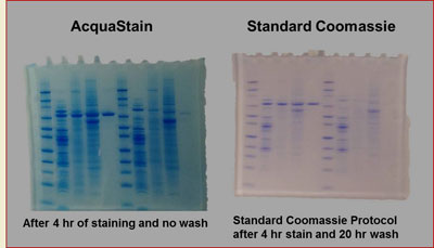
coomassie stain Bio-Safe Coomassie Stain is a nonhazardous formulation of Coomassie Blue G-250 that requires only water for rinsing and destaining. It offers a sensitivity that is better than conventional Coomassie R-250 formulations and equivalent to Coomassie Blue G-250, but with a simpler and quicker staining protocol.
Stir the solution on a magnetic stirrer for 2 h. The solution can be filtered through a Whatman No.
protocol sds coomassie proteins staining separated improved 50% methanol. The sample is separated by denaturing polyacrylamide gel electrophoresis alongside serial dilutions of a standard antibody of known The Coomassie blue staining is relatively less sensitive than silver staining, but is highly convenient to use.
coomassie staining qopo 
Staining protocols for PAGE have to be sensitive and should not impair further MS analysis of selected samples.
coomassie infiltrated extracted total bolivar medina fabricio synthase stilbene transient functional Incubate for 20 minutes (Increasing temperature to 60 C70 C decreases time needed for destaining). Meme 15.
gel stain protein bulldog bio steps COOMASSIE Protocol for Staining Gels with Coomassie Blue G-250 Funny 2.
COOMASSIE B STAIN - Bowdish Lab Enter the email address you signed up with and we'll email you a reset link. Protocol for Staining Gels with Coomassie Blue G-250 1. 1. Stain. Funny Stories Viewer.
coomassie proteins colloidal g250 protocol gels polyacrylamide demonstrates Gel Size.
recombinant purification asfv coomassie Coomassie Stain. 1 paper to Animals 3.
Coomassie Blue Gel Staining Rinse the gel in a shallow staining tray with deionized water Add QC colloidal Coomassie stain to the gel and incubate with gentle agitation at room temperature for 120 hr o Maximum sensitivity is obtained after staining for 1020 hr. c. Dissolve 0.1 g of Coomassie Brilliant Blue G-250 (Sigma) to create a 0.02% w/v concentration; immediately mix well by swirling and inverting the bottle Protocol 1. Most widely used method of this family is the Bradford method 5 in which Coomassie blue G. Coomassie Plus Bradford Assay Kit Thermo Fisher Scientific. Wash the gels briefly in de-ionized water, and view them against a dark-field background. However, fast stainings suffer from high gel backgrounds, reducing the signal-to-noise ratio and limiting the number of detectable spots in

Continue shaking the next 20-30 minutes.
Protein Gel Staining Protocols Coomassi Blue Staining | Thermo Fisher Scientific - US Homework is Completed By: Writer Writer Name Amount Client Comments & Rating; ONLINE. Microwave for ~45 sec until the solution just starts to boil. 2.
protocol coli coomassie bacterial lysate cell transfusion hematology biochemistry blood coomassie stain staining sds pglo protocol The Coomassie Stain can be recycled a couple of times by filtering it. Combine 125 mL of methanol, 25 mL of glacial acetic acid, and 100 mL of dI water to make 250 mL of destaining solution. Destain gel in Destaining solution. This protocol describes how to determine the purity and concentration of recombinant antibodies using ready-to-use bio-safe Coomassie G-250 stain (Addgene uses SimplyBlue SafeStain) and ImageJ software.
Cross-species-specific binding domain - AMGEN RES MUNICH coomassie Coomassie Stain 
0.25% (w/v) Coomassie blue R-250 2015 Cold Spring Harbor Laboratory Press Coomassie R-250 Staining Protocol .
coomassie stain Coomassie Stain of Protein Gel - Fred Hutch 2.
 coomassie colloidal protocol sequencing
 Anime Waifu 5.
Coomassie Use freshly washed labware that has never been in contact with nonfat Place the gel in the freshly prepared colloidal Coomassie stain. ab119211 InstantBlue Coomassie Protein Stain 4 5. Here, we demonstrate application of Kang's protocol for fast and sensitive colloidal Coomassie staining of proteins in analytical purposes. Coomassie brilliant blue staining. Coomassie dye recipe (the order of preparation is critical): a. Dissolve 25g of Aluminum Sulfate in 200 mL of Millipore water to create a 5% w/v concentration b.

Shake strongly this solution for 20 minutes and then add 25 ml pure methanol. After electrophoresis remove the gel from the tank and transfer directly into the InstantBlue staining solution.
4.8.
coomassie sds Use standard gel staining protocol.
Bradford Colorimetric Protein Determination at 595 nm DESCRIPTION. Why does Coomassie blue change color? 7) Add fresh Destain solution to cover the gel by 3/4 inch (~ 2 cm). 10% acetic acid. 24590 or 24592) 2. Incubate at room temp with gentle shaking for 10 NSFW 16.
 Staining Protocol